Registration
Landmark Detection in the Chest and Registration of Lung Surfaces in
Computed Tomography Scans
Margrit Betke, Harrison Hong, Chekema Prince, and Jane P. Ko
We developed an automated system for registering chest CT images
temporally. Our system detects anatomical landmarks in two CT scans
and matches them to obtain an initial alignment of the chests. The
lung surfaces are then segmented from the chests. We developed an
efficient algorithm to establish correspondences of lung surface
points. With this algorithm, lung surfaces are registered in an
iterative closest-point process, improving the initial alignment step
by step. We present a validation study that is based on registering
vessel branch points within the lungs. We applied our method to align
the lung surfaces of 10 pairs of chest CT scans and report a promising
registration performance.
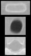
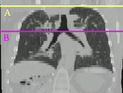
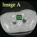
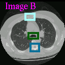
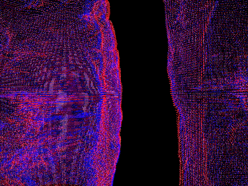
On the left, generic template images of the sternum, trachea,
and vertebra. Next, a coronal view of a chest CT scan. The yellow
line marks the most cranial image with visible lung (A), the purple
line the axial image at the carina (B). The trachea in image A
(green) is detected by correlation-based template matching using the
generic trachea template on the left. Sternum and vertebra in image B
(light and dark blue) are detected using their respective generic
templates. The trachea in image B (green) is found using a template
cropped online from the preceding axial image.
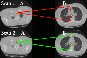
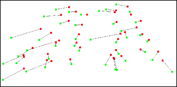
Initial landmark registration: Four points used for
registration are shown for each scan: the center of the trachea
cross-section in slice A and the centers of the cross-sections of
sternum, trachea, and vertebra in slice B in each study. The
landmarks in study 2 (green) are then be matched to the landmarks in
study 1 (red).
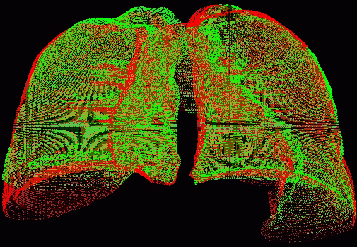
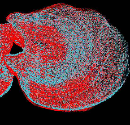
Registration results for high-resolution lung surfaces. The lung
surfaces are shown before and after registration. Zoomed-in views of
the lungs are given on the right. The lungs in scan 1 are shown in
red and in scan 2 prior to registration in green and after
registration in blue. The registration process shifted the surfaces
in scan 2 to the left and slightly rotated them to align with the
surfaces in scan 1
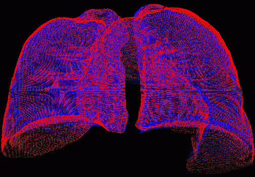
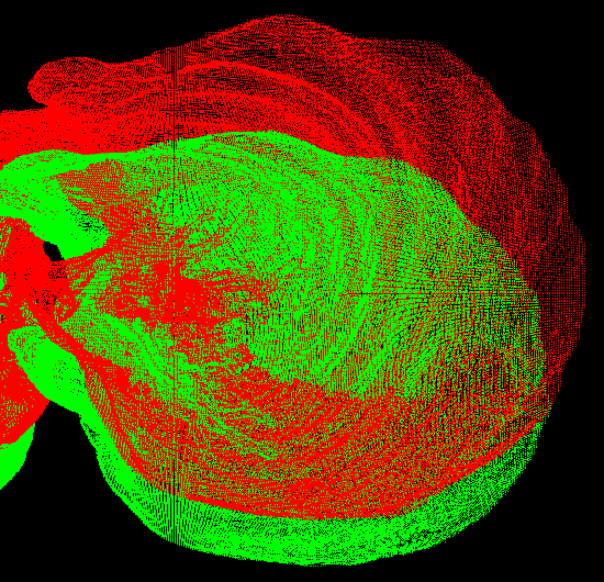
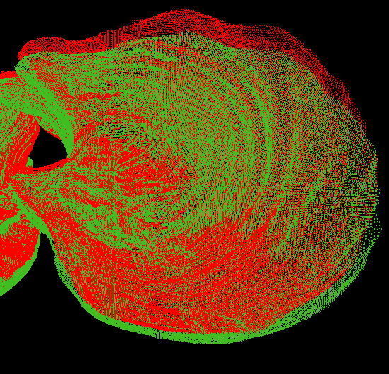
Top views of the right lung are given before any processing (left),
after the initial surface registration based on the landmark
registration parameters (middle), and after 25 iterations of the lung
surface registration (right). The surface in scan 1 is shown in red.
The surface in scan 2 is shown in green prior to registration and in
blue after registration.
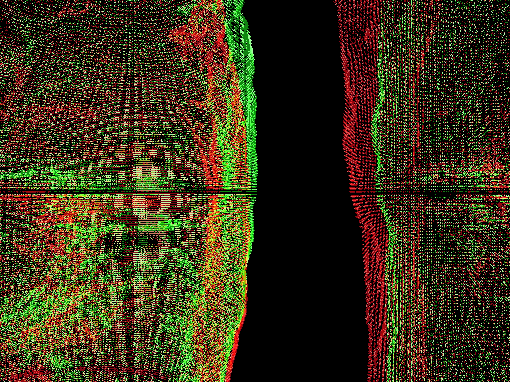
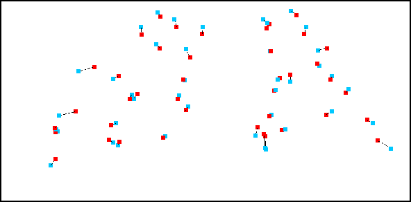
Validation of surface registration. The radiologist established point
correspondences of 42 vessel branching points in two CT studies of the
same patient. On top, a coronal view of these points in study 1 (red)
and study 2 (green) before registration. On the left, the points in
study 2 (blue) are aligned to the points in study 1 (red) by the
gold-standard rigid-body transformation that minimizes the sum of
squared differences (SSD) between the 42 point pairs. On the right,
the points in study 2 (blue) are aligned to the points in study 1
(red) using the rigid-body transformation that minimizes the SSD
between corresponding point pairs on the lung surfaces using our
registration algorithm. The translational difference between the two
transformations is less than 5 mm. The differences in Euler angles
are also small (2.1, 1.3, and 0.5 degrees).